How tumor cells spread to other parts of the body in skin cancer
- May 22, 2025
- 4 min read
First steps in metastasis formation clarified
Quick-change artists:
When melanoma cancer cells are attacked by the immune system, they activate specific genes and change their cell type. This reaction then inhibits T cells, among other things, making it easier for the skin cancer cells to colonize lymph nodes and form metastases, as researchers report in "Nature Cancer." They also identified proteins characteristic of these metastasis founder cells that may be suitable targets for therapy.

When a person develops cancer, it often doesn't stop at a single tumor. Over time, the cancer can spread and affect other parts of the body, even after the initial tumor has been removed. Whether a tumor forms such metastases depends on its genetics, but also on the patient's stress, diet, and treatment. Metastases cause diverse organ damage and, unlike the primary tumor, are often incurable. But how exactly do cancer cells colonize other organs?
According to one theory, cancer stem cells are drivers of metastasis. For example, degenerated, immature mammary gland cells from tumors attempt to form breast tissue in the liver or lungs. Although this attempt fails, the cancer stem cells still establish themselves in the foreign site and form new cancerous tumors there.
Skin Cancer as a Test Case
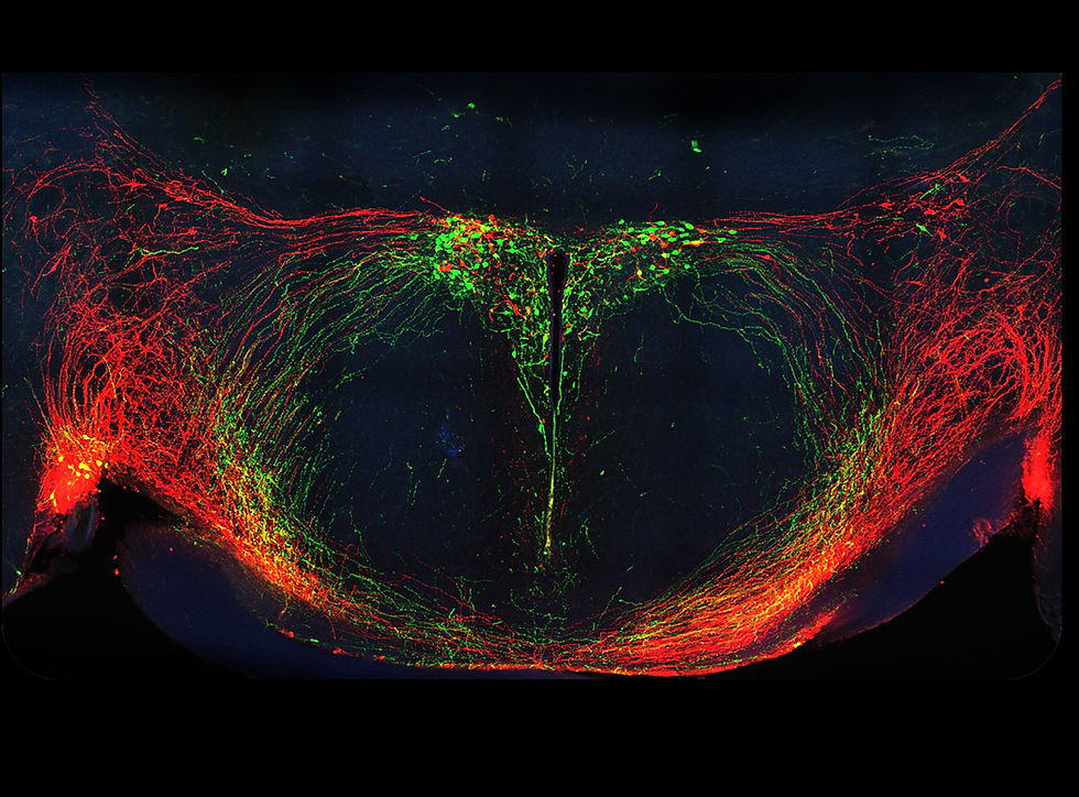
A team led by Severin Guetter from the University of Regensburg has now, for the first time, directly investigated this theory in cancer patients, rather than in animal models as previously done. To do so, the researchers compared biopsy samples from the lymph nodes of 492 patients with malignant melanoma, a type of black skin cancer, in stages I to III. About 40 percent of patients with metastases die from skin cancer.
The team used special dye markers to search the lymph node tissue for newly colonizing cancer cells from the melanoma. They then analyzed the properties of these cells.

Biomarkers for metastatic cancer cells
Guetter and his colleagues discovered that certain disseminated melanoma cells are associated with a significantly worse prognosis for cure and survival for patients. Even if the researchers found only one such cell among two million lymph node cells, this meant a worse course of disease for those affected than without this class of melanoma cells.
Further investigations showed that these cancer cells have the protein MCSP on their surface and produce at least one of the three proteins PMEL, MLANA, and DCT. The combination of these biomarkers allows for the unambiguous identification of metastatic melanoma cells.
Cancer cells switch on embryonic genes
Genetic analyses also revealed that these melanoma cells repeatedly alter their gene activity during their colonization in the lymph nodes, thus changing their appearance and metabolism. Among other things, the embryonic gene program, which is otherwise only active in melanocytes, is switched on. This causes these disseminated cancer cells to transition into a state similar to stem cells.
Through this reprogramming, the metastatic cancer cells develop into a cell type similar to the immature precursors of skin pigment cells. However, since the disseminated melanoma cells are not located in the skin but in the lymph node, "organ formation" fails, and metastases develop instead.
In contrast to classic cancer stem cells, the metastatic cancer cells in the tests responded dynamically to their microenvironment, the team emphasizes. This suggests that they are not static cancer stem cells, but rather a different, more flexible type of metastasis founder cells.

Battle with the immune system as a trigger
But what triggers this "rejuvenation" of the disseminated cancer cells? As Guetter and his colleagues discovered in further analyses, the human immune system is involved: The trigger for this development is apparently a previous battle between the melanoma cells and the immune system's T cells. These "killer cells" recognize cancer-typical characteristics on degenerated cells and attack them. However, the cancer cells that survive the T-cell attack subsequently activate the melanocyte gene program, as the tests showed.
In addition to MCSP production, part of this rejuvenating genomic program of the cancer cells also includes the production and release of proteins (CD155 and CD276) that suppress the immune system. This makes it easier for the disseminated cancer cells to establish themselves in the lymph nodes and form metastases, as the team explains. The larger these metastasis founder colonies become, the more proteins they release, and the more strongly this inhibits the T cells. The metastases can then grow unchecked, and the cancer cells they contain can once again alter their gene activity.
New Targets for Skin Cancer Therapy
The researchers have thus elucidated for the first time how the very first steps of metastasis formation in lymph nodes occur in melanoma. They assume that a similar process also occurs in metastases in other parts of the body. This knowledge could help in the future to treat skin cancer patients in such a way that metastasis formation is nipped in the bud.
A possible starting point here could be the MCSP protein, which appears to be carried by all dangerous metastasis founder cells but is rarely found in healthy body cells. Alternatively, the early immune response could be supported by T cells by targeting the proteins CD155 and CD276 to make it difficult for melanoma cells to establish a home. (Nature Cancer, 2025; doi: 10.1038/s43018-025-00963-w)
Source: University of Regensburg